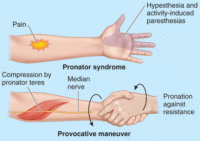


Ĭonservative treatment includes rest, modification of activities that exacerbate the symptoms, physical and occupational therapy, nonsteroidal anti-inflammatory medications, and local injections with corticosteroids or local anesthetic. Several studies with an ultrasound evaluation of median nerve between the humeral and ulnar heads of PT concluded that cross-sectional area of MN positively correlates with severity, duration of symptoms, and nerve conduction failure. Pronator teres syndrome, among other entrapments within the upper limb, also can be diagnosed by ultrasound and magnetic resonance imaging, but ultrasound is advantageous due to its dynamic character and lower cost. Some authors report abnormal electrodiagnostic findings in 10% of pronator teres syndrome only. If any of the median muscles are abnormal, other muscles innervated by the same myotomes as the proximal median muscles but supplied by a different nerve should be tested to exclude more proximal lesions within the brachial plexus or cervical roots. Electromyography (EMG) abnormalities occur in FPL and FDP to digits 2 and 3, less often in the FDS and APB, and only rarely in PT because compression at the site most often occurs distal to its innervation. Sensory and motor amplitudes are reduced more than conduction velocities, mostly in patients with severe and axonal symptoms. It is important that nerve conduction studies (NCS) are done in pronator teres syndrome to rule out other neuropathies, but they seldom show abnormalities. The anterior interosseous nerve (AIN) then branches from the MN about 5 to 8 cm distal to the medial epicondyle. Many individuals have additional fibrous brands within the two heads of the PT muscle. The absence of the ulnar head is rare (14%) and may reduce the risk of median nerve entrapment. Before the two heads unite, the median nerve passes between them in 74% to 82% of the cases, innervating both heads from C6-7 roots. They pass down to the forearm, form a common flexor tendon, and insert into the radial shaft. In the majority of cases (66%), it arises from unequal two heads: the larger humeral head from the upper part of the medial epicondyle and the smaller ulnar head from the coronoid process of the ulna. The PT muscle is named because of its action and shape it is a rounded muscle that pronates the forearm. Pronator teres syndrome (PTS), first described by Henrik Seyffarth in 1951, is caused by a compression of the median nerve (MN) by the pronator teres (PT) muscle in the forearm.


 0 kommentar(er)
